Virtual Family
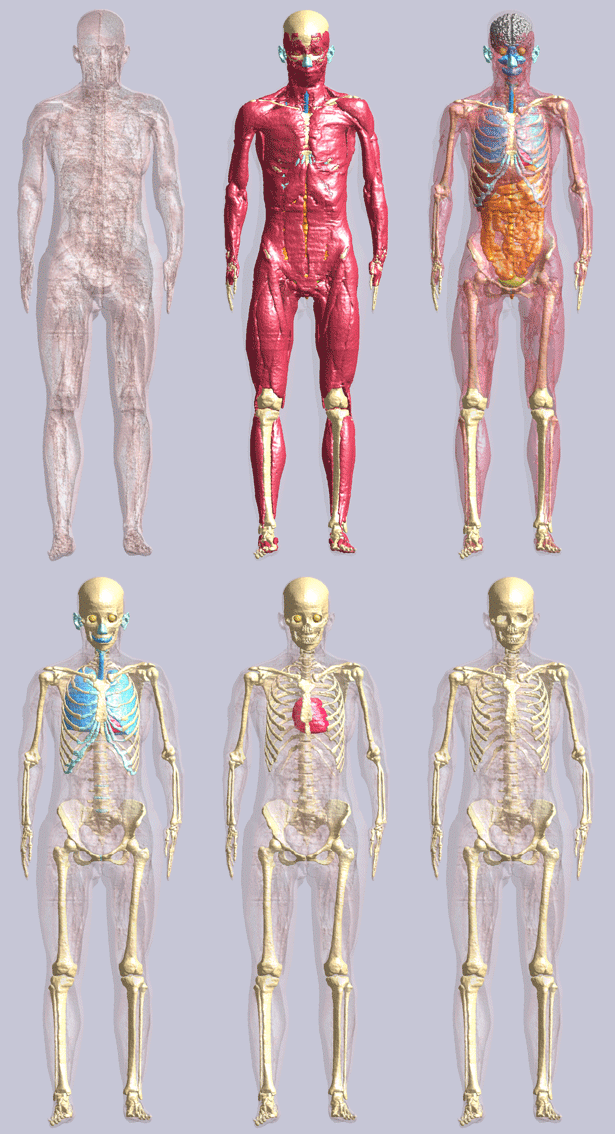
The Virtual Family1 (VF) is a set of four highly detailed, anatomically correct whole-body models of an adult male, an adult female, and two children. The VF project was carried out in collaboration between the U.S. Food and Drug Administration (FDA), the Foundation for Research on Information Technologies in Society (IT'IS Foundation, Zürich, Switzerland), Schmid & Partner Engineering AG (SPEAG, Zurich, Switzerland), the Hospital of the Friedrich-Alexander-University, Erlangen, Germany, and Siemens Medical Solutions, Erlangen, Germany. The Mobile Manufacturers Forum and the GSM Association also supported the development of the models, which were initially released in 2007. ZMT Zurich MedTech AG (ZMT, Zurich, Switzerland) and the IT'IS Foundation have sponsored the development of the newly released VF 2.0 models, with enhancements that improve spatial resolution, surface quality, and the effectiveness of the computer-aided design (CAD) handling.
The four VF models are based on high-resolution magnetic resonance imaging (MRI) data of healthy volunteers. Organs and tissues of the VF 2.0 models are represented by three-dimensional, highly detailed CAD objects without self-intersections and gaps. The CAD objects allow the models to be meshed at arbitrary resolutions without loss of small features.
Currently, the VF models are used for electromagnetic, thermal, acoustic, and computational fluid dynamics (CFD) simulations. Examples of applications of electromagnetic and thermal simulations are the assessment of the safety of active and passive medical implants in an MRI environment and the evaluation of the safety and efficacy of ablation devices2. Electromagnetic and thermal simulations have been performed on the entire set of VF models and additional models of children to calculate the whole-body averaged and local specific absorption rate (SAR) during exposure to 1.5 and 3T whole-body MRI coils3,4. These electromagnetic and thermal simulations have also allowed the evaluation of the safety of multi-channel transmit radio frequency whole-body MRI coils5. An example of application of electromagnetic and CFD simulations is the assessment of the applicability of the magneto-hemodynamic effect as a biomarker for cardiac output6. Acoustic simulations have been performed to assess the impact of the human anatomy on the focus location, shape, and intensity of ultrasound waves during focused ultrasound treatment7. As of the end of 2014, the VF was used in more than 120 medical device submissions to FDA and was cited more than 180 times in peer-reviewed literature. Recently the Virtual Population 3.0 became available8.
The following two VF models versions are available free of charge, except for handling fees:
VF 1.0: The VF 1.0 models include segmentation of approximately 80 high-resolution organs and tissues. The Virtual Family Tool can be used to discretize and export the CAD objects in a generic voxel-based format. All four VF 1.0 models and the Virtual Family Tool are provided free of charge (except for handling fees) to the scientific community for academic purposes only.
VF 2.0: The VF 2.0 models consist of simplified CAD files optimized for finite-element modeling in any third-party platform. These models are based on a new, high-end generation of VF models that have been re-segmented at finer resolution to afford a higher degree of precision and anatomical refinement, as well as improved structural continuity of approximately 300 organs and tissues. For the purpose of simplification, these structures are combined into 22 high-resolution tissues. The VF 2.0 models are available free of charge (except for handling fees) to everyone.
Virtual Family models
- DUKE: 34-year-old male
- ELLA: 26-year-old female
- BILLIE: 11-year-old female
- THELONIOUS: 6-year-old male
|  Ella |
|
|
Contact Information
Please address any questions or suggestions to Dr. Wolfgang Kainz at 301-661-7595 or [email protected].
References
- Christ A., Kainz W., Hahn E.G., Honegger K., Zefferer M., Neufeld E., Rascher W., Janka R., Bautz W., Chen J., Kiefer B., Schmitt P., Hollenbach H.P., Shen J.X., Oberle M., and Kuster N., “The Virtual Family - Development of Anatomical CAD Models of two Adults and two Children for Dosimetric Simulations”, Physics in Medicine and Biology, 55, N23–N38, 2010
- Cabot E., Lloyd T., Christ A., Kainz W., Douglas M., Stenzel G., Wedan S. and Kuster N., “Evaluation of the RF Heating of a Generic Deep Brain Stimulator Exposed in 1.5T Magnetic Resonance Scanners”, Bioelectromagnetics, 34(2):104-13, 2013
- Murbach M., Neufeld E., Capstick M., Kainz W., Brunner D., Samaras T., Pruessmann K., Kuster N., “Thermal Damage Tissue Models Analyzed for Different Whole-Body SAR and Scan Duration for Standard MR Body Coils”, Magnetic Resonance in Medicine, 2013
- Murbach M., Neufeld E., Kainz W., Pruessmann K, Kuster N., “Whole Body and Local RF Absorption in Human Models as a Function of Anatomy and Position within 1.5T MR Body Coil”, Magnetic Resonance in Medicine, 2013
- Neufeld E., Gosselin M.-C., Murbach M., Christ A., Cabot E. and Kuster N., “Analysis of the local worst-case SAR exposure caused by an MRI multi-transmit body coil in anatomical models of the human body”, Phys. Med. Biol. 56, 4649–4659, 2011
- Kyriakou A., Neufeld E., Szczerba D., Kainz W., Luechinger R., Kozerke S., McGregor R., Kuster N., “Patient-specific simulations and measurements of the magneto-hemodynamic effect in human primary vessels”, Physiol. Meas., 33(2):117-30, 2012
- Kyriakou, A., Neufeld, E., Werner, B., Paulides, M., Szekely, G. & Kuster, N., “A Review of Numerical and Experimental Compensation Techniques for Skull-Induced Phase Aberrations in Transcranial Focused Ultrasound”, International Journal of Hyperthermia, 30(1):36-46, 2014
- Gosselin M.-C., Neufeld E., Moser H., Huber E., Farcito S., Gerber L., Jedensjö M., Hilber I., Di Gennaro F., Lloyd B., Cherubini E., Szczerba D., Kainz W., Kuster N., “Development of a New Generation of High-Resolution Anatomical Models for Medical Device Evaluation: The Virtual Population 3.0”, Phys. Med. Biol. 59, 5287–5303, 2014
Disclaimer: The Food and Drug Administration (FDA) and the IT’IS Foundation assume no responsibility whatsoever for use of the Virtual Family by other parties and make no guarantees, expressed or implied, about the quality, reliability, or any other characteristic of the models. Further, use of the Virtual Family in no way implies endorsement by the FDA or confers any advantage in regulatory decisions. Version 2.0 of the Virtual Family is available free of charge from the IT'IS Foundation subject to a license agreement for protection of privacy. Any use of the Virtual Family is to be cited as stipulated in the license agreement. In addition, any derivative work shall bear a notice that it is sourced from the Virtual Family, and any modified versions shall additionally bear the notice that they have been modified. Version 2.0 of the Virtual Family and any derivative work or modified version thereof shall not be distributed to any third party whatsoever.


