Method for Fabrication of Microcalcifications for Insertion into Phantoms used to Evaluate X-ray Breast Imaging Systems
Catalog of Regulatory Science Tools to Help Assess New Medical Devices
Technical Description
This tool is a method for fabricating physical microcalcification simulants for insertion into breast phantoms for x-ray imaging with realistic chemical composition, x-ray attenuation, scatter properties, and adjustable density and sizes [1].
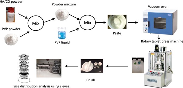
The preparation process is composed of seven steps, including cross-linking commercially available hydroxyapatite and calcium oxalate powders with polyvinylpyrrolidone (PVP) and mechanical compression [1].
These fabricated microcalcification simulants are intended to make the current breast phantoms more realistic for evaluating task-based detection performance of all breast imaging techniques, including x-ray transmission spectral imaging and x-ray coherent scatter computed tomography [1,2,3,4,5,6].
The method is described in detail in Fabrication of microcalcifications for insertion into phantoms used to evaluate x-ray breast imaging systems - IOPscience.
Figure 1. Schematic presentation of the method used in preparation of the phantom materials.
Intended Purpose
The microcalcifications simulants are intended to be inserted into breast phantoms for the evaluation of breast imaging technologies such as mammography, digital breast tomosynthesis, breast CT and spectral mammography.
The microcalcification phantoms can be used for objective, quantitative, task-based assessment of the detection and classification performance between the two microcalcification types typically found in breast cancer imaging.
Testing
The fabricated microcalcifications were evaluated by measuring their x-ray attenuation and scatter properties using x-ray spectroscopy and x-ray diffraction systems. Micro-CT was also used to evaluate the uniformity and reproducibility of the formulated tablets.
The microcalcification models' attenuation and scatter properties mimic the same properties of the real microcalcifications found in the breast. In addition, the micro-CT images confirm that the method is reproducible, and that appropriate density of the phantoms is retained after crushing to different sizes.
Several studies have used these microcalcifications to assess the task performance of microcalcification detection and classification [1,2,3,4,5,6].
Limitations
The proposed method is limited to microcalcification simulants for x-ray imaging modalities only.
Supporting Documentation
- Ghammraoui, B., Zidan, A., Alayoubi, A., Zidan, A., & Glick, S. J. (2021). Fabrication of microcalcifications for insertion into phantoms used to evaluate x-ray breast imaging systems. Biomedical physics & engineering express, 7(5), 10.1088/2057-1976/ac1c64. https://doi.org/10.1088/2057-1976/ac1c64
- Ikejimba, Salad, J., Graff, C. G., Goodsitt, M., Chan, H., Huang, H., Zhao, W., Ghammraoui, B., Lo, J. Y., & Glick, S. J. (2021). Assessment of task‐based performance from five clinical DBT systems using an anthropomorphic breast phantom. Medical Physics., 48(3), 1026–1038. https://doi.org/10.1002/mp.14568
- Ikejimba, Salad, J., Graff, C. G., Ghammraoui, B., Cheng, W., Lo, J. Y., & Glick, S. J. (2019). A four‐alternative forced choice (4AFC) methodology for evaluating microcalcification detection in clinical full‐field digital mammography (FFDM) and digital breast tomosynthesis (DBT) systems using an inkjet‐printed anthropomorphic phantom. Medical Physics., 46(9), 3883–3892. https://doi.org/10.1002/mp.13629
- Ghammraoui, B., Gkoumas, S., & Glick, S. J. (2021). Characterization of a GaAs photon-counting detector for mammography. Journal of medical imaging (Bellingham, Wash.), 8(3), 033504. https://doi.org/10.1117/1.JMI.8.3.033504
- Makeev, A., Rodal, G., Ghammraoui, B., Badal, A., & Glick, S. J. (2021). Exploring CNN potential in discriminating benign and malignant calcifications in conventional and dual-energy FFDM: simulations and experimental observations. Journal of medical imaging (Bellingham, Wash.), 8(3), 033501. https://doi.org/10.1117/1.JMI.8.3.033501
- Ghammraoui, B., Makeev, A., Zidan, A., Alayoubi, A., & Glick, S. J. (2019). Classification of breast microcalcifications using dual-energy mammography. Journal of medical imaging (Bellingham, Wash.), 6(1), 013502. https://doi.org/10.1117/1.JMI.6.1.013502
Contact
Tool Reference
In addition to citing relevant publications please reference the use of this tool using DOI: 10.5281/zenodo.8383595

