amyotrophic lateral sclerosis
How Double-Stranded RNA Protects the Brain Against Infection While Making Damaging Neuroinflammation More Likely
Posted on by Lawrence Tabak, D.D.S., Ph.D.

When you get a run-of-the-mill viral infection, after a few days of symptoms your immune system typically fends off the bug, and you’ll make a full recovery. In rare cases, a virus can infect the brain. This can lead to much bigger problems, including cognitive impairments known as “brain fog,” other neuropsychiatric symptoms, potentially irreversible brain damage, or even death. For this reason, the brain, more than other parts of the body, relies heavily on immune responses that can control viral infections immediately.
Now some intriguing findings from an NIH-funded team reported in Science Immunology help to explain how the brain is protected against infections.1 However, the findings also highlight a serious downside: these same mechanisms that protect the brain also leave it especially vulnerable to damaging levels of neuroinflammation.
The new findings may help to explain what goes on in the brains of people with a wide range of neurodegenerative conditions, including amyotrophic lateral sclerosis (ALS) and Alzheimer’s disease. They also point to promising targets for developing treatments that might turn inflammatory immune responses in the brain up or down, as desired, to treat these and other serious conditions.
How does it work? The key is double-stranded RNA (dsRNA).
RNA molecules are readouts of genetic information in DNA that carry instructions for building the proteins that carry out various cell functions. RNA molecules in our cells are most often in single-stranded or short dsRNA form. In contrast, lengthy dsRNAs are a hallmark of viruses. When a virus invades our cells, our immune system’s first line of defense can sense those long viral dsRNAs and trigger a response.
But it turns out that dsRNAs aren’t unique to viruses, as the new study highlights. The researchers, led by Tyler Dorrity and Heegwon Shin, both members of Hachung Chung’s lab at Columbia University Irving Medical Center, New York, found that human neurons—even when they’re normal and healthy—also have exceptionally high levels of long dsRNAs.
Their lab studies in cells and tissues show that these dsRNAs in neurons can trigger an inflammatory immune response just as they do in viruses. By manipulating neurons in a way that cut back on the number of dsRNAs, they found they could lower the innate immune response. However, cells with fewer dsRNAs also showed greater susceptibility to infection with Zika viruses and herpes simplex virus, which can produce a form of viral encephalitis.
The researchers also knew from earlier studies that people with a rare, inherited condition called Aicardi-Goutières syndrome (AGS), which primarily affects the brain and immune system, carry a mutation that causes their cells to lack an enzyme needed to edit dsRNAs. As a result, neurons carrying this mutation have so many dsRNAs that it is toxic.
They went on to show that they could shift this dynamic by altering levels of two other proteins that bind RNA. The proteins normally encourage dsRNA formation in the brain. When the researchers deleted these RNA-binding proteins from the AGS neurons, those neurons made fewer long dsRNAs, which in turn protected them from the inflammatory immune responses and allowed them to survive longer. As expected, however, those cells also were more susceptible to viral infection.
The findings show how this tricky balance between susceptibility to infection and inflammation in the brain works in both health and disease. It also leads to the tantalizing suggestion that treatments targeting these various players or others in the same pathways may offer new ways of treating brain infections or neuroinflammatory conditions, by boosting or dampening dsRNA levels and the associated immune responses. As a next step, the researchers report that they’re pursuing studies to explore the role of dsRNA-triggered immune responses in ALS and Alzheimer’s, as well as in neuropsychiatric symptoms sometimes seen in people with lupus.
References:
[1] TJ Dorrity TJ, et al. Long 3’UTRs predispose neurons to inflammation by promoting immunostimulatory double-stranded RNA formation. Science Immunology DOI: 10.1126/sciimmunol.adg2979 (2023).
NIH Support: National Institute of Neurological Disorders and Stroke, National Institute of Allergy and Infectious Diseases, National Institute of General Medical Sciences
Finding Beauty in Cell Stress
Posted on by Dr. Francis Collins
Most stressful situations that we experience in daily life aren’t ones that we’d choose to repeat. But the cellular stress response captured in this video is certainly worth repeating a few times, so you can track what happens when two cancer cells get hit with stressors.
In this movie of two highly stressed osteosarcoma cells, you first see the appearance of many droplet-like structures (green). This is followed by a second set of droplets (magenta) and, finally, the fusion of both types of droplets.
These droplets are composed of fluorescently labeled stress-response proteins, either G3BP or UBQLN2 (Ubiquilin-2). Each protein is undergoing a fascinating process, called phase separation, in which a non-membrane bound compartment of the cytoplasm emerges and constrains the motion of proteins within it. Subsequently, the proteins fuse with like proteins to form larger droplets, in much the same way that raindrops merge on a car’s windshield.
Julia Riley, an undergraduate student in the NIH-supported lab of Heidi Hehnly and lab of Carlos Castañeda, Syracuse University, NY, shot this movie using the sophisticated tools of fluorescence microscopy. It’s the next installment in our series featuring winners of the 2019 Green Fluorescent Protein Image and Video Contest, sponsored by the American Society for Cell Biology. The contest honors the discovery of green fluorescent protein (GFP), which—together with a rainbow of other fluorescent proteins—has enabled researchers to visualize proteins and their dynamic activities inside cells for the last 25 years.
Riley and colleagues suspect that, in this case, phase separation is a protective measure that allows proteins to wall themselves off from the rest of the cell during stressful conditions. In this way, the proteins can create new functional units within the cell. The researchers are working to learn much more about what this interesting behavior entails as a basic organizing principle in the cell and how it works.
Even more intriguing is that similar stress-responding proteins are commonly altered in people with the devastating neurologic condition known as amyotrophic lateral sclerosis (ALS). ALS is a group of rare neurological diseases that involve the progressive deterioration of neurons responsible for voluntary movements such as chewing, walking, and talking. There’s been the suggestion that these phase separation droplets may seed the formation of the larger protein aggregates that accumulate in the motor neurons of people with this debilitating and fatal condition.
Castañeda and Hehnly, working with J. Paul Taylor at St. Jude Children’s Research Hospital, Memphis, earlier reported that Ubiquilin-2 forms stress-induced droplets in multiple cell types [1]. More recently, they showed that mutations in Ubiquilin-2 have been linked to ALS changes in the way that the protein undergoes phase separation in a test tube [2].
While the proteins in this award-winning video aren’t mutant forms, Riley is now working on the sequel, featuring versions of the Ubiquilin-2 protein that you’d find in some people with ALS. She hopes to capture how those mutations might produce a different movie and what that might mean for understanding ALS.
References:
[1] Ubiquitin Modulates Liquid-Liquid Phase Separation of UBQLN2 via Disruption of Multivalent Interactions. Dao TP, Kolaitis R-M, Kim HJ, O’Donovan K, Martyniak B, Colicino E, Hehnly H, Taylor JP, Castañeda CA. Molecular Cell. 2018 Mar 15;69(6):965-978.e6.
[2] ALS-Linked Mutations Affect UBQLN2 Oligomerization and Phase Separation in a Position- and Amino Acid-Dependent Manner. Dao TP, Martyniak B, Canning AJ, Lei Y, Colicino EG, Cosgrove MS, Hehnly H, Castañeda CA. Structure. 2019 Jun 4;27(6):937-951.e5.
Links:
Amyotrophic Lateral Sclerosis (ALS) (National Institute of Neurological Disorders and Stroke/NIH)
Castañeda Lab (Syracuse University, NY)
Hehnly Lab (Syracuse University)
Green Fluorescent Protein Image and Video Contest (American Society for Cell Biology, Bethesda, MD)
2008 Nobel Prize in Chemistry (Nobel Foundation, Stockholm, Sweden)
NIH Support: National Institute of General Medical Sciences
Making Personalized Blood-Brain Barriers in a Dish
Posted on by Dr. Francis Collins

The blood-brain barrier, or BBB, is a dense sheet of cells that surrounds most of the brain’s blood vessels. The BBB’s tiny gaps let vital small molecules, such as oxygen and water, diffuse from the bloodstream into the brain while helping to keep out larger, impermeable foreign substances that don’t belong there.
But in people with certain neurological disorders—such as amyotrophic lateral sclerosis (ALS) and Huntington’s disease—abnormalities in this barrier may block the entry of biomolecules essential to healthy brain activity. The BBB also makes it difficult for needed therapies to reach their target in the brain.
To help look for solutions to these and other problems, researchers can now grow human blood-brain barriers on a chip like the one pictured above. The high-magnification image reveals some of the BBB’s cellular parts. There are endothelial-like cells (magenta), which are similar to those that line the small vessels surrounding the brain. In close association are supportive brain cells known as astrocytes (green), which help to regulate blood flow.
While similar organ chips have been created before, what sets apart this new BBB chip is its use of induced pluripotent stem cell (iPSC) technology combined with advanced chip engineering. The iPSCs, derived in this case from blood samples, make it possible to produce a living model of anyone’s unique BBB on demand.
The researchers, led by Clive Svendsen, Cedars-Sinai, Los Angeles, first use a biochemical recipe to coax a person’s white blood cells to become iPSCs. At this point, the iPSCs are capable of producing any other cell type. But the Svendsen team follows two different recipes to direct those iPSCs to differentiate into endothelial and neural cells needed to model the BBB.
Also making this BBB platform unique is its use of a sophisticated microfluidic chip, produced by Boston-based Emulate, Inc. The chip mimics conditions inside the human body, allowing the blood-brain barrier to function much as it would in a person.
The channels enable researchers to flow cerebral spinal fluid (CSF) through one side and blood through the other to create the fully functional model tissue. The BBB chips also show electrical resistance and permeability just as would be expected in a person. The model BBBs are even able to block the entry of certain drugs!
As described in Cell Stem Cell, the researchers have already created BBB chips using iPSCs from a person with Huntington’s disease and another from an individual with a rare congenital disorder called Allan-Herndon-Dudley syndrome, an inherited disorder of brain development.
In the near term, his team has plans to model ALS and Parkinson’s disease on the BBB chips. Because these chips hold the promise of modeling the human BBB more precisely than animal models, they may accelerate studies of potentially promising new drugs. Svendsen suggests that individuals with neurological conditions might one day have their own BBB chips made on demand to help in selecting the best-available therapeutic options for them. Now that’s a future we’d all like to see.
Reference:
[1] Human iPSC-Derived Blood-Brain Barrier Chips Enable Disease Modeling and Personalized Medicine Applications. Vatine GD, Barrile R, Workman MJ, Sances S, Barriga BK, Rahnama M, Barthakur S, Kasendra M, Lucchesi C, Kerns J, Wen N, Spivia WR, Chen Z, Van Eyk J, Svendsen CN. Cell Stem Cell. 2019 Jun 6;24(6):995-1005.e6.
Links:
Tissue Chip for Drug Screening (National Center for Advancing Translational Sciences/NIH)
Stem Cell Information (NIH)
Svendsen Lab (Cedars-Sinai, Los Angeles)
NIH Support: National Institute of Neurological Disorders and Stroke; National Center for Advancing Translational Sciences
A Tribute to Two Amazing Scientists
Posted on by Dr. Francis Collins
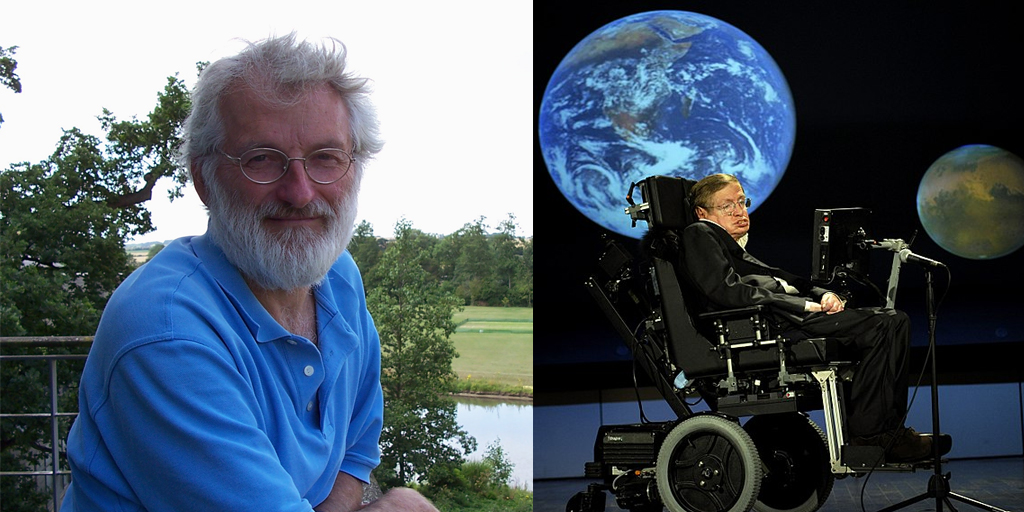
Caption: Sir John Sulston (left) and Stephen Hawking (right)
Credit: Jane Gitschier, PLoS; Paul Alers, NASA
Over the past couple of weeks, we’ve lost two legendary scientists who made major contributions to our world: Sir John Sulston and Stephen Hawking. Although they worked in very different areas of science—biology and physics—both have left us with an enduring legacy through their brilliant work that unlocked fundamental mysteries of life and the universe.
I had the privilege of working closely with John as part of the international Human Genome Project (HGP), a historic endeavor that successfully produced the first reference sequence of the human genetic blueprint nearly 15 years ago, in April 2003. As founding director of the Sanger Centre (now the Sanger Institute) in Cambridge, England, John oversaw the British contributions to this publicly funded effort. Throughout our many planning meetings and sometimes stormy weekly conference calls about progress of this intense and all-consuming enterprise, John stood out for his keen intellect and high ethical standards.
Creative Minds: Can Diseased Cells Help to Make Their Own Drugs?
Posted on by Dr. Francis Collins

Matthew Disney grew up in a large family in Baltimore in the 1980s. While his mother worked nights, Disney and his younger brother often tagged along with their father in these pre-Internet days on calls to fix the microfilm machines used to view important records at hospitals, banks, and other places of business. Watching his father take apart the machines made Disney want to work with his hands one day. Seeing his father work tirelessly for the sake of his family also made him want to help others.
Disney found a profession that satisfied both requirements when he fell in love with chemistry as an undergraduate at the University of Maryland, College Park. Now a chemistry professor at The Scripps Research Institute, Jupiter, FL, Disney is applying his hands and brains to develop a treatment strategy that aims to control the progression of a long list of devastating disorders that includes Huntington’s disease, amyotrophic lateral sclerosis (ALS), and various forms of muscular dystrophy.
The 30 or so health conditions on Disney’s list have something in common. They are caused by genetic glitches in which repetitive DNA letters (CAGCAGCAG, for example) in transcribed regions of the genome cause some of the body’s cells and tissues to produce unwieldy messenger RNA molecules that interfere with normal cellular activities, either by binding other intracellular components or serving as templates for the production of toxic proteins.
The diseases on Disney’s list also have often been considered “undruggable,” in part because the compounds capable of disabling the lengthy, disease-causing RNA molecules are generally too large to cross cell membranes. Disney has found an ingenious way around that problem [1]. Instead of delivering the finished drug, he delivers smaller building blocks. He then uses the cell and its own machinery, including the very aberrant RNA molecules he aims to target, as his drug factory to produce those larger compounds.
Disney has received an NIH Director’s 2015 Pioneer Award to develop this innovative drug-delivery strategy further. He will apply his investigational approach initially to treat a common form of muscular dystrophy, first using human cells in culture and then in animal models. Once he gets that working well, he’ll move on to other conditions including ALS.
What’s appealing about Disney’s approach is that it makes it possible to treat disease-affected cells without affecting healthy cells. That’s because his drugs can only be assembled into their active forms in cells after they are templated by those aberrant RNA molecules.
Interestingly, Disney never intended to study human diseases. His lab was set up to study the structure and function of RNA molecules and their interactions with other small molecules. In the process, he stumbled across a small molecule that targets an RNA implicated in a rare form of muscular dystrophy. His niece also has a rare incurable disease, and Disney saw a chance to make a difference for others like her. It’s a healthy reminder that the pursuit of basic scientific questions often can lead to new and unexpectedly important medical discoveries that have the potential to touch the lives of many.
Reference:
[1] A toxic RNA catalyzes the in cellulo synthesis of its own inhibitor. Rzuczek SG, Park H, Disney MD. Angew Chem Int Ed Engl. 2014 Oct 6;53(41):10956-10959.
Links:
Disney Lab (The Scripps Research Institute, Jupiter, FL)
Disney NIH Project Information (NIH RePORTER)
NIH Director’s Pioneer Award Program
NIH Support: Common Fund; National Institute of Neurological Disorders and Stroke