BioArt 2014
Snapshots of Life: New Target for Herpes Treatment?
Posted on by Dr. Francis Collins
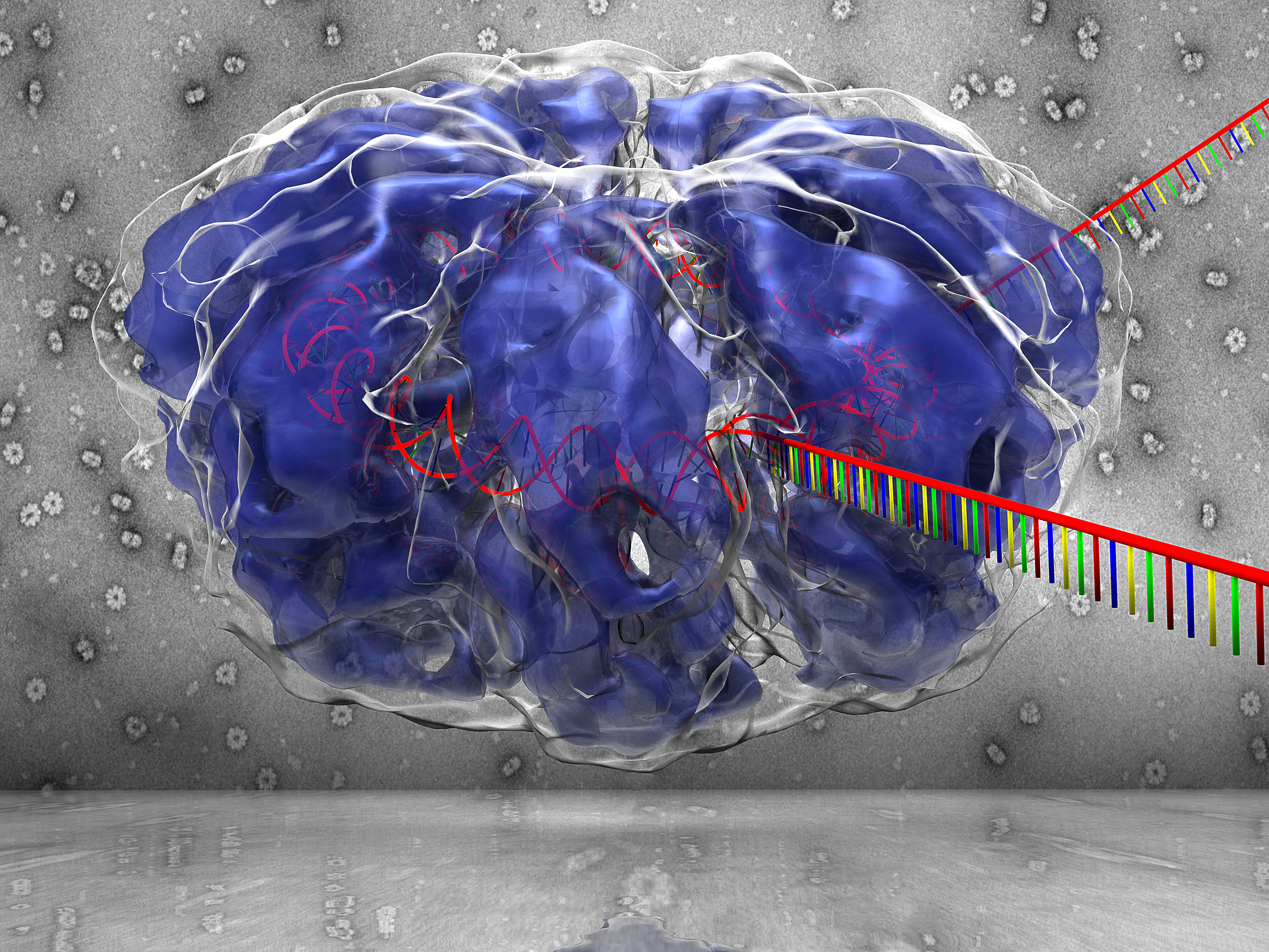 Something about this image reminds me of that wacky and infectious old song: “It was a one-eyed, one-horned, flyin’ purple people eater …” Of course, this purple blob isn’t a people eater, but it does happen to be infectious. What you see here is a 3D rendering of a protein that the herpes simplex virus 1 (HSV-1)—one of two herpes viruses that cause genital herpes and cold sores—depends upon to infect human cells.
When a cell is infected with HSV-1, the virus inserts its DNA into human cells, periodically coming out of dormancy to make more copies of itself. However, errors sometimes occur when the DNA is replicated. When that happens, an HSV-1 protein, dubbed infected cell protein 8 (ICP8), stitches broken pieces of DNA back together. That’s what you see depicted in this schematic, which shows two single strands of DNA (red with multicolor bases) entering an ICP8 complex (purplish blue) to be reannealed into DNA’s familiar double-stranded helix (red).
Snapshots of Life: A Colorful Look Inside the Retina
Posted on by Dr. Francis Collins

Credit: Amy Robinson, Alex Norton, William Silversmith, Jinseop Kim, Kisuk Lee, Aleks Zlasteski, Matt Green, Matthew Balkam, Rachel Prentki, Marissa Sorek, Celia David, Devon Jones, and Doug Bland, Massachusetts Institute of Technology, Cambridge, MA; Sebastian Seung, Princeton University, Princeton, NJ
This eerie scene might bring back memories of the computer-generated alien war machines from Steven Spielberg’s War of the Worlds thriller. But what you’re seeing is a computer-generated depiction of a quite different world—the world inside the retina, the light-sensitive tissue that lines the back of the eye. The stilt-legged “creatures” are actually ganglion nerve cells, and what appears to be their long “noses” are fibers that will eventually converge to form the optic nerve that relays visual signals to the brain. The dense, multi-colored mat near the bottom of the image is a region where the ganglia and other types of retinal cells interact to convey visual information.
What I find particularly interesting about this image is that it was produced through the joint efforts of people who played EyeWire, an internet crowdsourcing game developed in the lab of computational neuroscientist Sebastian Seung, now at Princeton University in New Jersey. Seung and his colleagues created EyeWire using a series of high-resolution microscopic images of the mouse retina, which were digitized into 3D cubes containing dense skeins of branching nerve fibers. It’s at this point where the crowdsourcing came in. Online gamers—most of whom aren’t scientists— volunteered for a challenge that involved mapping the 3D structure of individual nerve cells within these 3D cubes. Players literally colored-in the interiors of the cells and progressively traced their long extensions across the image to distinguish them from their neighbors. Sounds easy, but the branches are exceedingly thin and difficult to follow.
Snapshots of Life: The Biological Basis of Hearing
Posted on by Dr. Francis Collins

Credit: Peter Barr-Gillespie and Kateri Spinelli, Oregon Health & Science University, Portland
Did you know that chickens have ears? Well, here’s the evidence—you’re looking at a micrograph of sensory hair cells that make up the inner ear of Gallus gallus domesticus, otherwise known as the domestic chicken. Protruding from each hair cell is a tall bundle of stiff appendages, called stereocilia, that capture vibrations and enable the chicken to hear everything from grain being poured into a feeder to the footsteps of a wily fox. The flatter area is occupied by supporting cells, which have recently been shown to have the capacity to regenerate damaged or destroyed hair cells.
Peter Barr-Gillespie and Kateri Spinelli of Oregon Health & Science University, Portland used a scanning electron microscope to capture this image—one of the winners of the Federation of American Societies for Experimental Biology’s 2014 BioArt competition—while studying how these cells convert sound waves into brain waves. It is generally known that sound waves cause the stereocilia on each hair cell to oscillate in concert. These vibrating stereocilia trigger electrical changes in the hair cells, which then send signals to the brain. Barr-Gillespie’s group focuses on the actual molecules that build the stereocilia and translate the vibrations into brain signals.
Cool Videos: A Look Inside a Mouse’s Lung
Posted on by Dr. Francis Collins

If you have ever wondered what it is like to be an oxygen molecule inhaled through the lungs, here is your chance to find out! In this movie, we take a fantastic voyage through the slippery airways of the adult mouse lung.
We begin at the top in the main pipeline, called the bronchus, just below the trachea and wind through a system of increasingly narrow tubes. As you zoom through the airways, take note of the cilia (seen as goldish streaks); these tiny, hair-like structures move dust, germs, and mucus from smaller air passages to larger ones. Our quick trip concludes with a look into the alveoli — the air sacs where oxygen is delivered to red blood cells and carbon dioxide is removed and exhaled.
Cool Videos: HIV in Action
Posted on by Dr. Francis Collins

There aren’t too many molecular biologists who have spent a 3-month stint in Hollywood. But Janet Iwasa is not your average molecular biologist. After earning her PhD in 2006, she took a break from the lab to take a crash course in animation techniques at the Gnomon School of Visual Effects.
While her classmates produced lots of cool footage worthy of the silver screen, Iwasa wanted to learn how to depict in colorful 3D action, some of the complex molecular processes that are so difficult to convey using static 2D illustration. Among her creations is this 2-minute, rough-draft animation showing how the human immunodeficiency virus (HIV) recognizes and infects a type of immune cell known as a T cell.
Snapshots of Life: Lost Connections in Pompe Disease
Posted on by Dr. Francis Collins
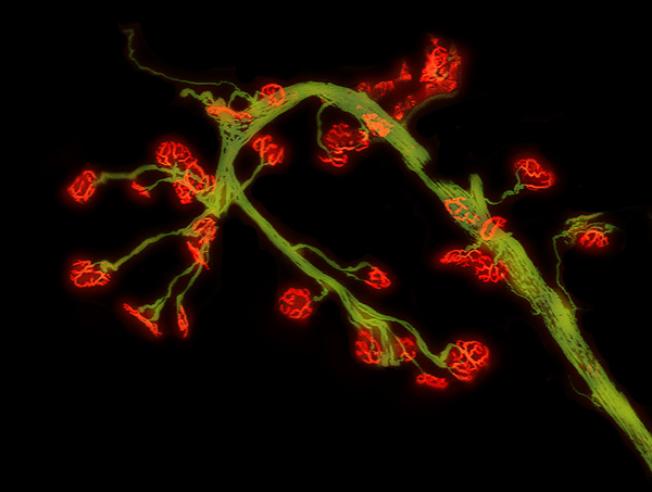
Caption: Abnormal connections between leg muscle fibers (red) and nerves (green) in Pompe disease.
Credit: Darin J. Falk, A. Gary Todd, Robin Yoon, and Barry J. Byrne, University of Florida, Gainesville
Mistletoe? Holly? Not exactly. This seemingly festive image is a micrograph of nerve cells (green) and nerve-muscle junctions (red) in a mouse model of Pompe disease. Such images are helping researchers learn more about this rare form of muscular dystrophy, providing valuable clues in the ongoing search for better treatments and cures.
People with Pompe disease lack an enzyme that cells depend on to break down a stored sugar, known as glycogen, into smaller glucose molecules that can be readily used for energy. Without enough of this enzyme, called acid alpha-glucosidase (GAA), glycogen can accumulate destructively in the liver, heart, and skeletal muscles, making it increasingly difficult to walk, eat, and even breathe.