Early Independence Award
Creative Minds: Searching for Solutions to Chronic Infection
Posted on by Dr. Francis Collins
If you or a loved one has ever struggled with a bacterial infection that seemed to have gone away with antibiotic treatment, but then came back again, you’ll probably be interested to learn about the work of Kyle Allison. What sometimes happens when a person has an infection—for instance, a staph infection of the skin—is that antibiotics kill off the vast majority of bacteria, but a small fraction remain alive. After antibiotic treatment ends, those lurking bacterial “persisters” begin to multiply, and the person develops a chronic infection that may be very difficult and costly to eliminate.
Unlike antibiotic-resistant superbugs, bacterial persisters don’t possess any specific genetic mutations that protect them against the killing power of one particular medication or another. Rather, the survival of these bacteria depends upon their ability to enter a dormant state that allows them to hang on in the face of antibiotic treatment. It isn’t clear exactly how the bugs do it, and that’s where Kyle’s work comes in.
LabTV: Curious about the Aging Brain
Posted on by Dr. Francis Collins
This LabTV video takes us to the West Coast to meet Saul Villeda, a creative young researcher who’s exploring ways to reduce the effects of aging on the human brain. Thanks to a 2012 NIH Director’s Early Independence award, Villeda set up his own lab at the University of California, San Francisco to study how age-related immune changes may affect the ability of brain cells to regenerate. By figuring out exactly what’s going on, Villeda and his team hope to devise ways to counteract such changes, possibly preventing or even reversing the cognitive declines that all too often come with age.
Villeda is the first person in his family to become a scientist. His parents immigrated to the United States from Guatemala, settled into a working-class neighborhood in Pasadena, CA, and enrolled their kids in public schools. While he was growing up, Villeda says he’d never even heard of a Ph.D. and thought all doctors were M.D.’s who wore stethoscopes. But he did have a keen mind and a strong sense of curiosity—gifts that helped him become the valedictorian of his high school class and find his calling in science. Villeda went on to earn an undergraduate degree in physiological science from the University of California, Los Angeles and a Ph.D. in neurosciences from Stanford University Medical School, Palo Alto, CA, as well as to publish his research findings in several influential scientific journals.
Creative Minds: Michael Angelo’s Art
Posted on by Dr. Francis Collins

Caption: The location and abundance of six proteins—e-cadherin (green), vimentin (blue), actin (red), estrogen receptor, progesterone receptor, and Ki67—found in breast cancer cells are seen in this multiplexed ion beam image. Cells positive for estrogen receptor a, progesterone receptor, and Ki-67 appear yellow; cells expressing estrogen receptor a and the progesterone receptor appear aqua.
Credit: Michael Angelo
The artistic masterpiece above, reminiscent of a stained glass window, is the work of Michael Angelo—no, not the famous 16th Century Italian artist, but a 21st Century physician-scientist who’s out to develop a better way of looking at what’s going on inside solid tumors. Called multiplexed ion beam imaging (MIBI), Angelo’s experimental method may someday give clinicians the power to analyze up to 100 different proteins in a single tumor sample.
In this image, Angelo used MIBI to analyze a human breast tumor sample for nine proteins simultaneously—each protein stained with an antibody tagged with a metal reporter. Six of the nine proteins are illustrated here. The subpopulation of cells that are positive for three proteins often used to guide breast cancer treatment (estrogen receptor a, progesterone receptor, Ki-67) have yellow nuclei, while aqua marks the nuclei of another group of cells that’s positive for only two of the proteins (estrogen receptor a, progesterone receptor). In the membrane and cytoplasmic regions of the cell, red indicates actin, blue indicates vimentin, which is a protein associated with highly aggressive tumors, and the green is E-cadherin, which is expressed at lower levels in rapidly growing tumors than in less aggressive ones. Taken together, such “multi-dimensional” information on the types and amounts of proteins in a patient’s tumor sample may give oncologists a clearer idea of how quickly that tumor is growing and which types of treatments may work best for that particular patient. It also shows dramatically how much heterogeneity is present in a group of breast cancer cells that would have appeared identical by less sophisticated methods.
Creative Minds: A Baby’s Eye View of Language Development
Posted on by Dr. Francis Collins
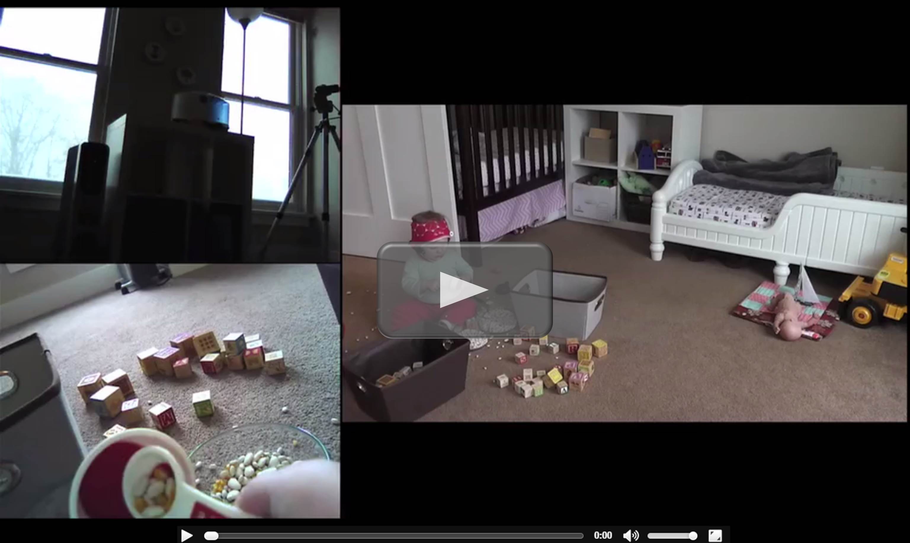
 If you are a fan of wildlife shows, you’ve probably seen those tiny video cameras rigged to animals in the wild that provide a sneak peek into their secret domains. But not all research cams are mounted on creatures with fur, feathers, or fins. One of NIH’s 2014 Early Independence Award winners has developed a baby-friendly, head-mounted camera system (shown above) that captures the world from an infant’s perspective and explores one of our most human, but still imperfectly understood, traits: language.
Elika Bergelson, a young researcher at the University of Rochester in New York, wants to know exactly how and when infants acquire the ability to understand spoken words. Using innovative camera gear and other investigative tools, she hopes to refine current thinking about the natural timeline for language acquisition. Bergelson also hopes her work will pay off in a firmer theoretical foundation to help clinicians assess children with poor verbal skills or with neurodevelopmental conditions that impair information processing, such as autism spectrum disorders.
Creative Minds: Tackling Chemotherapy Resistance
Posted on by Dr. Francis Collins
For many young scientists, nothing can equal the chance to have a lab of one’s own. Still, it often takes considerable time to get there. To help creative minds cut to the chase sooner, the NIH Director’s Early Independence Awards this year will enable 17 outstanding young researchers to skip post-doctoral training and begin running their own labs immediately.
Today, I’d like to tell you about one of these creative minds. His name is Aaron Meyer, a cell signaling expert at the Massachusetts Institute of Technology in Cambridge, and his research project will take aim at one the biggest challenges in cancer treatment: chemotherapy resistance.
Cool Videos: Myotonic Dystrophy
Posted on by Dr. Francis Collins
Today, I’d like to share a video that tells the inspirational story of two young Massachusetts Institute of Technology (MIT) researchers who are taking aim at a genetic disease that has touched both of their lives. Called myotonic dystrophy (DM), the disease is the most common form of muscular dystrophy in adults and causes a wide variety of health problems—including muscle wasting and weakness, irregular heartbeats, and profound fatigue.
If you’d like a few more details before or after watching these scientists’ video, here’s their description of their work: “Eric Wang started his lab at MIT in 2013 through receiving an NIH Early Independence Award. Learn about the path that led him to study myotonic dystrophy, a disease that affects his family. Eric’s team of researchers includes Ona McConnell, an avid field hockey goalie who is affected by myotonic dystrophy herself. Determined to make a difference, Eric and Ona hope to inspire others in their efforts to better understand and treat this disease.”
Links:
Creative Minds: Targeting Cancer with Lasers and Nanoballoons
Posted on by Dr. Francis Collins
When most people think about cancer treatments, what typically come to mind are the side effects of traditional chemotherapy: cardiac, liver, and renal toxicity; hair loss; nausea; fatigue—just to name a few. These side effects occur because the cancer drugs damage not just cancer cells, but healthy cells as well. “Targeted” cancer therapy, on the other hand, is designed to target just the cancer cells. Some targeted therapies achieve this because they only attack cells with a particular molecular signature; others are directed to the cancer by physical means. Today, I’d like to introduce you to a researcher who’s developing a targeted drug delivery strategy that uses lasers and light activated drug delivery to fight cancer.
Jonathan Lovell, a Canadian-born researcher at the State University of New York at Buffalo (UB) and recipient of the NIH Director’s Early Independence Award, has designed unique nanosized spherical pods—1/1000 the diameter of a human hair—that open when light shines on them and snap shut in the dark. Lovell will fill these pods, also known as liposomes—hollow fat droplets—with anti-cancer drugs. He’ll then inject them into the body, where they’ll circulate, safely and silently: until they’re activated. When Lovell shines a red laser on the tumor, the light triggers the balloons to open and deliver a blast of the drug—only where it is needed. (Red light penetrates human tissue better than other colors.) It’s a terrific example of how bioengineering can bring fresh solutions to longstanding medical challenges.
Creative Minds: Lighting Up Memory
Posted on by Dr. Francis Collins
 One of the most debilitating, and heartbreaking, consequences of Alzheimer’s disease is the way it slowly robs people of their memories. Unfortunately, we don’t yet have a cure for Alzheimer’s, let alone a good understanding of exactly how this disease destroys memory skills. That’s why, in this first post in my series highlighting some of the awardees in NIH Common Fund’s High-Risk, High-Reward Research Program, I’m excited to introduce a young scientist who’s using some cool technology to tackle this formidable challenge: Christine Ann Denny.
A winner of a 2013 NIH Director’s Early Independence Awards (often called the “skip-the-postdoc” award), Denny has developed a technique to label the cells that encode individual memories in the brains of mice. That’s right: she tags the nerve cells that build these memories, the neurons, with a fluorescent molecule that glows.
Forbes 30 Under 30 Highlights NIH Stars
Posted on by Dr. Francis Collins
Wow! Seeing this new Forbes list just made my day! It’s inspiring to glimpse the up and coming young minds who will be shaping tomorrow’s science. But what makes me particularly proud is that four of them—Mitchell Guttman, Gregory Sonnenberg, Adam de la Zerda, and Daniela Witten— are recent recipients of the NIH Director’s Early Independence Award—a “skip the postdoc” grant that allows young minds to unleash their creativity, talent, independence, and drive.
Here’s a quick taste of just what makes these grantees so noteworthy. Guttman, an assistant professor at Caltech, is studying a new type of gene that regulates embryonic development. Gregory Sonnenburg, an immunologist at University of Pennsylvania, studies the role of beneficial bacteria in the gut and why the immune system sometimes turns against these friends. Daniela Witten, assistant professor at the University of Washington, is creating machine learning programs that massage vast amounts of data into useful and actionable knowledge—one example is personalized cancer therapy. Adam de la Zerda, assistant professor at Stanford, is using nanotechnology to understand cancer and age-related macular degeneration.
Another exceptional advocate for medical research tops the Forbes’ list of 30 under 30. Josh Sommer, a young man who was diagnosed with a rare cancer called chordoma when he was 18, is someone I have had the pleasure of encouraging and mentoring. Josh now runs the Chordoma Foundation that has raised $2.5 million and supports research in 11 labs.
All of these young scientists are amazing, and I look forward to seeing all the wonderful innovative work they do.
With a healthy dose of tongue in cheek, I’m happy to announce that the AARP just chose me as one of the “50 over 50” influential leaders—so I guess there’s also hope at the other end of the spectrum.
Happy holidays, everyone!


