hypothalmus
‘Tis the Season for Good Cheer
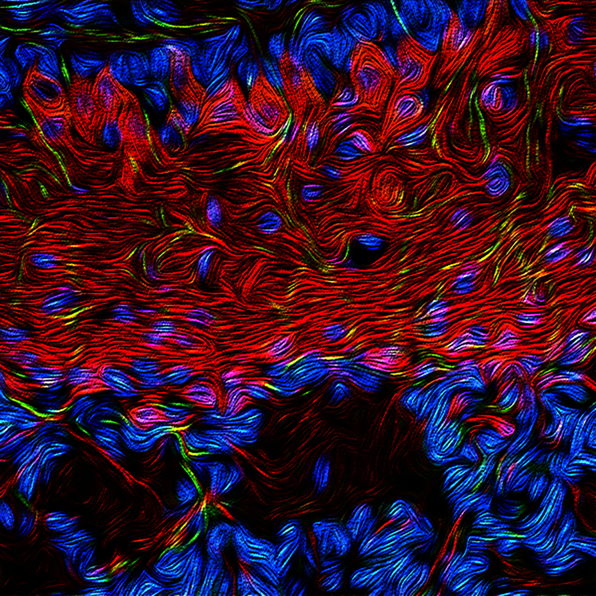
Posted on by Dr. Francis Collins

Credit: Oka lab, California Institute of Technology, Pasadena
Whether it’s Rockefeller Center, the White House, or somewhere else across the land, ‘tis the season to gather with neighbors for a communal holiday tree-lighting ceremony. But this festive image has more do with those cups of cider in everyone’s hands than admiring the perfect Douglas fir. What looks like lights and branches are actually components of a high-resolution map from a part of the brain that controls thirst.
The map, drawn up from mouse studies, shows that when thirst arises, neurons activate a gene called c-fos (red)—lighting up the tree—indicating it’s time for a drink. In response, other neurons (green) direct additional parts of the brain to compensate by managing internal water levels. In a mouse that’s no longer thirsty, the tree would look almost all green.
This wiring map comes from a part of the brain called the hypothalamus, which is best known for its role in hunger, thirst, and energy balance. Thanks to powerful molecular tools from NIH’s Brain Research through Advancing Innovative Technologies (BRAIN) Initiative, Yuki Oka of the California Institute of Technology, Pasadena, and his team were able to draw detailed maps of the tree-shaped region, called the median preoptic nucleus (MnPO).
Using a technique called optogenetics, Oka’s team, led by Vineet Augustine, could selectively turn on genes in the MnPO [1]. By doing so, they could control a mouse’s thirst and trace the precise control pathways responsible for drinking or not.
This holiday season, as you gather with loved ones, take a moment to savor the beautiful complexity of biology and the gift of human health. Happy holidays to all of you, and peace and joy into the new year!
Reference:
[1] Hierarchical neural architecture underlying thirst regulation. Augustine V, Gokce SK, Lee S, Wang B, Davidson TJ, Reimann F, Gribble F, Deisseroth K, Lois C, Oka Y. Nature. 2018 Mar 8;555(7695):204-209.
Links:
Oka Lab, California Institute of Technology, Pasadena
The BRAIN Initiative (NIH)
NIH Support: National Institute of Neurological Disorders and Stroke
Unraveling the Biocircuitry of Obesity
Posted on by Dr. Francis Collins

Caption: Mouse neurons (purple), with their nuclei (blue) and primary cilia (green).
Credit: Yi Wang, Vaisse Lab, UCSF
Obesity involves the complex interplay of diet, lifestyle, genetics, and even the bacteria living in the gut. But there are other less-appreciated factors that are likely involved, and a new NIH-supported study suggests one that you probably never would have imagined: antenna-like sensory projections on brain cells.
The study in mice, published in the journal Nature Genetics [1], suggests these neuronal projections, called primary cilia, are a key part of a known “hunger circuit,” which receives signals from other parts of the body to control appetite. The researchers add important evidence in mouse studies showing that changes in the primary cilia can produce a short circuit, impairing the brain’s ability to regulate appetite and leading to overeating and obesity.
Snapshots of Life: Making Sense of Smell
Posted on by Dr. Francis Collins
Credit: Jeremy McIntyre, University of Florida College of Medicine, Gainesville
You’ve probably learned the hard way about how the grocery list can go out the window when you go shopping on an empty stomach. Part of the reason is that our sense of smell intensifies when we’re hungry, making the aroma of freshly baked cookies, fried chicken, and other tempting goodies even more noticeable. And this beautiful micrograph helps to provide a biological explanation for this phenomenon.
The image, which looks like something that Van Gogh might have painted, shows a thick mesh of neurons in a small cross section of a mouse’s olfactory bulb, a structure located in the forebrain of all vertebrates (including humans!) that processes input about odors detected by the nose. Here, you see specialized neurons called mitral cells (red) that can receive signals from the hypothalamus, a brain region known for its role in hunger and energy balance. Also fluorescently labeled are receptors that detect acetylcholine signals from the brain (green) and the nuclei of all cells in the olfactory bulb (blue).