induced Pluripotent Stem cells
Studying Color Vision in a Dish
Posted on by Dr. Francis Collins

Credit: Eldred et al., Science
Researchers can now grow miniature versions of the human retina—the light-sensitive tissue at the back of the eye—right in a lab dish. While most “retina-in-a-dish” research is focused on finding cures for potentially blinding diseases, these organoids are also providing new insights into color vision.
Our ability to view the world in all of its rich and varied colors starts with the retina’s light-absorbing cone cells. In this image of a retinal organoid, you see cone cells (blue and green). Those labelled with blue produce a visual pigment that allows us to see the color blue, while those labelled green make visual pigments that let us see green or red. The cells that are labeled with red show the highly sensitive rod cells, which aren’t involved in color vision, but are very important for detecting motion and seeing at night.
Snapshots of Life: Growing Mini-Brains in a Dish
Posted on by Dr. Francis Collins

Credit: Collin Edington and Iris Lee, Department of Biomedical Engineering, MIT
Something pretty incredible happens—both visually and scientifically—when researchers spread neural stem cells onto a gel-like matrix in a lab dish and wait to see what happens. Gradually, the cells differentiate and self-assemble to form cohesive organoids that resemble miniature brains!
In this image of a mini-brain organoid, the center consists of a clump of neuronal bodies (magenta), surrounded by an intricate network of branching extensions (green) through which these cells relay information. Scattered throughout the mini-brain are star-shaped astrocytes (red) that serve as support cells.
Creative Minds: Mapping the Biocircuitry of Schizophrenia and Bipolar Disorder
Posted on by Dr. Francis Collins

Bruce Yankner
As a graduate student in the 1980s, Bruce Yankner wondered what if cancer-causing genes switched on in non-dividing neurons of the brain. Rather than form a tumor, would those genes cause neurons to degenerate? To explore such what-ifs, Yankner spent his days tinkering with neural cells, using viruses to insert various mutant genes and study their effects. In a stroke of luck, one of Yankner’s insertions encoded a precursor to a protein called amyloid. Those experiments and later ones from Yankner’s own lab showed definitively that high concentrations of amyloid, as found in the brains of people with Alzheimer’s disease, are toxic to neural cells [1].
The discovery set Yankner on a career path to study normal changes in the aging human brain and their connection to neurodegenerative diseases. At Harvard Medical School, Boston, Yankner and his colleague George Church are now recipients of an NIH Director’s 2016 Transformative Research Award to apply what they’ve learned about the aging brain to study changes in the brains of younger people with schizophrenia and bipolar disorder, two poorly understood psychiatric disorders.
Creative Minds: A Transcriptional “Periodic Table” of Human Neurons
Posted on by Dr. Francis Collins

Caption: Mouse fibroblasts converted into induced neuronal cells, showing neuronal appendages (red), nuclei (blue) and the neural protein tau (yellow).
Credit: Kristin Baldwin, Scripps Research Institute, La Jolla, CA
Writers have The Elements of Style, chemists have the periodic table, and biomedical researchers could soon have a comprehensive reference on how to make neurons in a dish. Kristin Baldwin of the Scripps Research Institute, La Jolla, CA, has received a 2016 NIH Director’s Pioneer Award to begin drafting an online resource that will provide other researchers the information they need to reprogram mature human skin cells reproducibly into a variety of neurons that closely resemble those found in the brain and nervous system.
These lab-grown neurons could be used to improve our understanding of basic human biology and to develop better models for studying Alzheimer’s disease, autism, and a wide range of other neurological conditions. Such questions have been extremely difficult to explore in mice and other animal models because they have shorter lifespans and different brain structures than humans.
Regenerative Medicine: Making Blood Stem Cells in the Lab
Posted on by Dr. Francis Collins

Caption: Arrow in first panel points to an endothelial cell induced to become hematopoietic stem cell (HSC). Second and third panels show the expansion of HSCs over time.
Credit: Raphael Lis, Weill Cornell Medicine, New York, NY
Bone marrow transplants offer a way to cure leukemia, sickle cell disease, and a variety of other life-threatening blood disorders.There are two major problems, however: One is many patients don’t have a well-matched donor to provide the marrow needed to reconstitute their blood with healthy cells. Another is even with a well-matched donor, rejection or graft versus host disease can occur, and lifelong immunosuppression may be needed.
A much more powerful option would be to develop a means for every patient to serve as their own bone marrow donor. To address this challenge, researchers have been trying to develop reliable, lab-based methods for making the vital, blood-producing component of bone marrow: hematopoietic stem cells (HSCs).
Two new studies by NIH-funded research teams bring us closer to achieving this feat. In the first study, researchers developed a biochemical “recipe” to produce HSC-like cells from human induced pluripotent stem cells (iPSCs), which were derived from mature skin cells. In the second, researchers employed another approach to convert mature mouse endothelial cells, which line the inside of blood vessels, directly into self-renewing HSCs. When these HSCs were transplanted into mice, they fully reconstituted the animals’ blood systems with healthy red and white blood cells.
Regenerative Medicine: The Promise and Peril
Posted on by Dr. Francis Collins
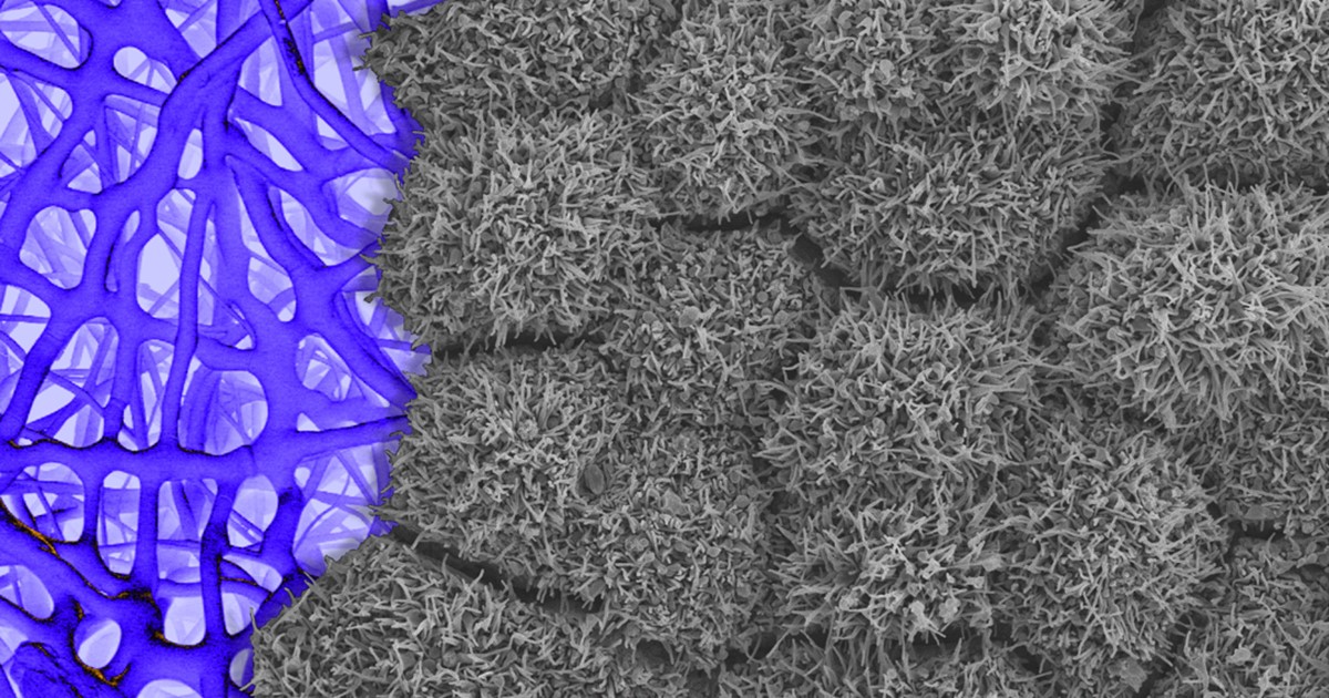
Caption: Scanning electron micrograph of iPSC-derived retinal pigment epithelial cells growing on a nanofiber scaffold (blue).
Credit: Sheldon Miller, Arvydas Maminishkis, Robert Fariss, and Kapil Bharti, National Eye Institute/NIH
Stem cells derived from a person’s own body have the potential to replace tissue damaged by a wide array of diseases. Now, two reports published in the New England Journal of Medicine highlight the promise—and the peril—of this rapidly advancing area of regenerative medicine. Both groups took aim at the same disorder: age-related macular degeneration (AMD), a common, progressive form of vision loss. Unfortunately for several patients, the results couldn’t have been more different.
In the first case, researchers in Japan took cells from the skin of a female volunteer with AMD and used them to create induced pluripotent stem cells (iPSCs) in the lab. Those iPSCs were coaxed into differentiating into cells that closely resemble those found near the macula, a tiny area in the center of the eye’s retina that is damaged in AMD. The lab-grown tissue, made of retinal pigment epithelial cells, was then transplanted into one of the woman’s eyes. While there was hope that there might be actual visual improvement, the main goal of this first in human clinical research project was to assess safety. The patient’s vision remained stable in the treated eye, no adverse events occurred, and the transplanted cells remained viable for more than a year.
Exciting stuff, but, as the second report shows, it is imperative that all human tests of regenerative approaches be designed and carried out with the utmost care and scientific rigor. In that instance, three elderly women with AMD each paid $5,000 to a Florida clinic to be injected in both eyes with a slurry of cells, including stem cells isolated from their own abdominal fat. The sad result? All of the women suffered severe and irreversible vision loss that left them legally or, in one case, completely blind.
Progress Toward Stem Cell Treatment for Diabetes
Posted on by Dr. Francis Collins

Caption: Insulin-containing pancreatic beta cells (green) derived from human stem cells. The red cells are producing another metabolic hormone, glucagon, that regulates blood glucose levels. Blue indicates cell nuclei.
Credit: The Salk Institute for Biological Studies, La Jolla, CA
In people with type 1 diabetes, the immune system kills off insulin-producing beta cells of the pancreas needed to control the amount of glucose in their bloodstream. As a result, they must monitor their blood glucose often and take replacement doses of insulin to keep it under control. Transplantation of donated pancreatic islets—tissue that contains beta cells—holds some promise as a therapy or even a cure for type 1 diabetes. However, such donor islets are in notoriously short supply [1]. Recent advances in stem cell research have raised hopes of one day generating an essentially unlimited supply of replacement beta cells perfectly matched to the patient to avoid transplant rejection.
A couple of years ago, researchers took a major step toward this goal by coaxing induced pluripotent stem cells (iPSCs), which are made from mature human cells, to differentiate into cells that closely resembled beta cells. But a few things were troublesome. The process was long and difficult, and the iPSC-derived cells were not quite as good at sensing glucose and secreting insulin as cells in a healthy person. They also looked and, in some ways, acted like beta cells, but were unable to mature fully in the lab. Now, an NIH-funded team has succeeded in finding an additional switch that enables iPSC-derived beta cells to mature and produce insulin in a dish—a significant step toward moving this work closer to the clinical applications that many diabetics have wanted.
LabTV: Curious About Parkinson’s Disease
Posted on by Dr. Francis Collins

When the young scientist featured in this LabTV video first learned about induced pluripotent stem (iPS) cells a few years ago as an undergrad, he thought it would be cool if he could someday work with this innovative technology. Today, as a graduate student, Kinsley Belle is part of a research team that’s using iPS cells on a routine basis to gain a deeper understanding of Parkinson’s disease.
Derived from genetically reprogrammed skin cells or white blood cells, iPS cells have the potential to develop into many different types of cells, providing scientists with a powerful tool to model a wide variety of diseases in laboratory dishes. At the University of Miami’s John P. Hussman Institute for Human Genomics, Belle and his colleagues are taking advantage of an iPS model of Parkinson’s disease to explore its molecular roots. Their goal? To use that information to develop better treatments or maybe even a cure for the neurodegenerative disorder that affects at least a half-million Americans.
Bioengineering: Big Potential in Tiny 3D Heart Chambers
Posted on by Dr. Francis Collins

Caption: Heart microchamber generated from human iPS cells; cardiomyocytes (red), myofibroblasts (green), cell nuclei (blue)
Credit: Zhen Ma, University of California, Berkeley
The adult human heart is about the size of a large fist, divided into four chambers that beat in precise harmony about 100,000 times a day to circulate blood throughout the body. That’s a very dynamic system, and also a very challenging one to study in real-time in the lab. Understanding how the heart forms within developing human embryos is another formidable challenge. So, you can see why researchers are excited by the creation of tiny, 3D heart chambers with the ability to exist (see image above) and even beat (see video below) in a lab dish, or as scientists say “in vitro.”

Credit: Zhen Ma et al., Nature Communications
To achieve this feat, an NIH-funded team from University of California, Berkeley, and Gladstone Institute of Cardiovascular Disease, San Francisco turned to human induced pluripotent stem (iPS) cell technology. The resulting heart chambers may be miniscule—measuring no more than a couple of hair-widths across—but they hold huge potential for everything from improving understanding of cardiac development to speeding drug toxicity screening.
Next Page