model
A New View of the 3D Genome
Posted on by Dr. Francis Collins
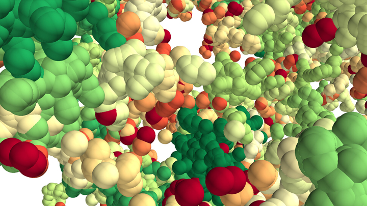
This lush panoply of color might stir up daydreams of getting away to explore a tropical rain forest. But what you see here is a new model that’s enabling researchers to explore something equally amazing: how a string of DNA that measures 6 feet long can be packed into the microscopic nucleus of a human cell. Fitting that much DNA in a nucleus is like fitting a thread the length of the Empire State building underneath your fingernail!
Scientists have known for a while that that the answer lies in how DNA is folded onto spool-like complexes called chromatin, but many details of the process still remain to be worked out. Recently, an NIH-funded team, led by Vadim Backman and Igal Szleifer, Northwestern University, Evanston, IL, developed this new model of chromatin folding by pairing sophisticated mathematical modeling and optical imaging.In a study published in the journal Science Advances [1], the team found that chromatin is folded into a variety of tree-like domains along a chromatin backbone, which they liken to an aggregation of trees growing from the forest floor. The colorful spheres you see above represent trees of varying sizes.
Earlier models of chromatin folding had suggested that DNA folds into regular and orderly fibers. In the new study, the Northwestern researchers used their own specially designed Partial Wave Spectroscopic microscope. This high-powered system, coupled with electron imaging, allowed them to peer deep inside living cells to “sense” real-time alterations in chromatin packing. What makes their new view on chromatin so interesting is it suggests our DNA is packaged in a way that’s much more disorderly and unpredictable than initially thought.
As Backman notes, it is reasonable to assume that a forest would be filled with trees of varying sizes and shapes. But you couldn’t predict the exact location of each tree or its particular size and configuration. The same appears to be true of these tree-like structures within chromatin. Their precise location and size vary, seemingly unpredictably, from cell to cell.
This apparently random DNA packing structure might seem surprising given chromatin’s importance in influencing the expression and function of our genes. But the researchers think such variability likely has its advantages.
Here’s the idea: If all of our cells responded to stressful conditions (such as heat or a toxic exposure) in exactly the same way and that way happened to be suboptimal, the whole tissue or organ might fail. But if differences in chromatin structure lead each cell to respond somewhat differently to the same stimulus, then some cells might be more likely to survive or even thrive under the stress. It’s a built-in way for cells to hedge their bets.
These new findings offer a fundamentally new three-dimensional view of the human genome. They might also inspire innovative strategies to understand and fight cancer, as well as other diseases. And, while most of us probably won’t be venturing off into the rain forest anytime soon, this work does give us all something to think about next time we’re enjoying the great outdoors in our own neck of the woods.
Reference:
[1] Physical and data structure of 3D genome. Huang K, Li Y, Shim AR, Virk RKA, Agrawal V, Eshein A, Nap RJ, Almassalha LM, Backman V, Szleifer I. Sci Adv. 2020 Jan 10;6(2):eaay4055.
Links:
Deoxyribonucleic Acid (DNA) (National Human Genome Research Institute/NIH)
4D Nucleome (Common Fund/NIH)
Vadim Backman (Northwestern University, Evanston, IL)
Igal Szleifer (Northwestern University, Evanston, IL)
NIH Support: National Cancer Institute
3D Printing the Novel Coronavirus
Posted on by Dr. Francis Collins

The coronavirus disease 2019 (COVID-19) pandemic has truly been an all-hands-on-deck moment for the nation. Among the responders are many with NIH affiliations, who are lending their expertise to deploy new and emerging technologies to address myriad research challenges. That’s certainly the case for the dedicated team from the National Institute of Allergy and Infectious Diseases (NIAID) at the NIH 3D Print Exchange (3DPX), Rockville, MD.
A remarkable example of the team’s work is this 3D-printed physical model of SARS-CoV-2, the novel coronavirus that causes COVID-19. This model shows the viral surface (blue) and the spike proteins studded proportionally to the right size and shape. These proteins are essential for SARS-CoV-2 to attach to human cells and infect them. Here, the spike proteins are represented in their open, active form (orange) that’s capable of attaching to a human cell, as well as in their closed, inactive form (red).
The model is about 5 inches in diameter. It takes more than 5 hours to print using an “ink” of thin layers of a gypsum plaster-based powder fused with a colored binder solution. When completed, the plaster model is coated in epoxy for strength and a glossy, ceramic-like finish. For these models, NIAID uses commercial-grade, full-color 3D printers. However, the same 3D files can be used in any type of 3D printer, including “desktop” models available on the consumer market.
Darrell Hurt and Meghan McCarthy lead the 3DPX team. Kristen Browne, Phil Cruz, and Victor Starr Kramer, the team members who helped to produce this remarkable model, created it as part of a collaboration with the imaging team at NIAID’s Rocky Mountain Laboratories (RML), Hamilton, MT.
The RML’s Electron Microscopy Unit captured the microscopic 3D images of the virus, which was cultured from one of the first COVID-19 patients in the country. The unit handed off these and other data to its in-house visual specialist to convert into a preliminary 3D model. The model was then forwarded to the 3DPX team in Maryland to colorize and optimize in preparation for 3D printing.
This model is especially unique because it’s based exclusively on SARS-CoV-2 data. For example, the model is assembled from data showing that the virus is frequently oval, not perfectly round. The spike proteins also aren’t evenly spaced, but pop up more randomly from the surface. Another nice feature of 3D printing is the models can be constantly updated to incorporate the latest structural discoveries.
That’s why 3D models are such an excellent teaching tools to share among scientists and the public. Folks can hold the plaster virus and closely examine its structure. In fact, the team recently printed out a model and delivered it to me for exactly this educational purpose.
In addition to this complete model, the researchers also are populating the online 3D print exchange with atomic-level structures of the various SARS-CoV-2 proteins that have been deposited by researchers around the world into protein and electron microscopy databanks. The number of these structures and plans currently stands at well over 100—and counting.
As impressive as this modeling work is, 3DPX has found yet another essential way to aid in the COVID-19 fight. In March, the Food and Drug Administration (FDA) announced a public-private partnership with the NIH 3D Print Exchange, Department of Veterans Affairs (VA) Innovation Ecosystem, and the non-profit America Makes, Youngstown, OH [1]. The partnership will develop a curated collection of designs for 3D-printable personal protective equipment (PPE), as well as other necessary medical devices that are in short supply due to the COVID-19 pandemic.
You can explore the partnership’s growing collection of COVID-19-related medical supplies online. And, if you happen to have a 3D printer handy, you could even try making them for yourself.
Reference:
[1] FDA Efforts to Connect Manufacturers and Health Care Entities: The FDA, Department of Veterans Affairs, National Institutes of Health, and America Makes Form a COVID-19 response Public-Private Partnership (Food and Drug Administration)
Links:
Coronavirus (COVID-19) (NIH)
NIH 3D Print Exchange (National Institute of Allergy and Infectious Diseases/NIH, Rockville, MD)
Rocky Mountain Laboratories (NIAID/NIH, Hamilton, MT)
Department of Veterans Affairs (VA) Innovation Ecosystem (Washington, D.C.)
America Makes (Youngstown, OH)
NIH Support: National Institute of Allergy and Infectious Diseases