scanning electron micrograph
Snapshots of Life: Stronger Than It Looks
Posted on by Dr. Francis Collins
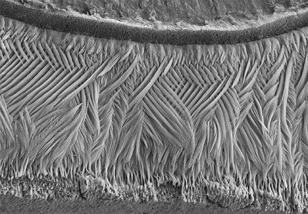
Credit: Olivier Duverger and Maria I. Morasso, National Institute of Arthritis and Musculoskeletal and Skin Diseases, NIH
If you went out and asked folks what they’re seeing in this picture, most would probably guess an elegantly woven basket, or a soft, downy feather. But what this scanning electron micrograph actually shows isn’t at all soft: it is the hardest substance in the mammalian body—tooth enamel!
This exquisitely detailed image—a winner of the Federation of American Societies for Experimental Biology’s 2015 BioArt competition—was generated by Olivier Duverger and Maria Morasso of NIH’s National Institute of Arthritis and Musculoskeletal and Skin Diseases. Before placing a sample of mouse dental enamel under the microscope, they treated it briefly with acid in order to reveal how the tissue’s mineralized rods are interwoven in a manner that gives teeth both strength and flexibility.
Snapshots of Life: Sore Throat as Art
Posted on by Dr. Francis Collins

Credit: Vincent A. Fischetti, The Rockefeller University
Most parents and kids wouldn’t consider strep throat the subject of high art. But the judges of the Federation of American Societies for Experimental Biology’s 2013 BioArt competition think it is. In this silver-toned scanning electron micrograph, you can see hundreds of tiny spheres—bacteria called Group A streptococci—attached to a human pharyngeal (throat) cells grown in a lab dish. These bacteria are responsible for a very nasty type of pharyngeal inflammation commonly known as strep throat. Strep infections are usually treated with antibiotics; left untreated, they can lead to rheumatic fever, rheumatic heart disease, and even kidney disease.